


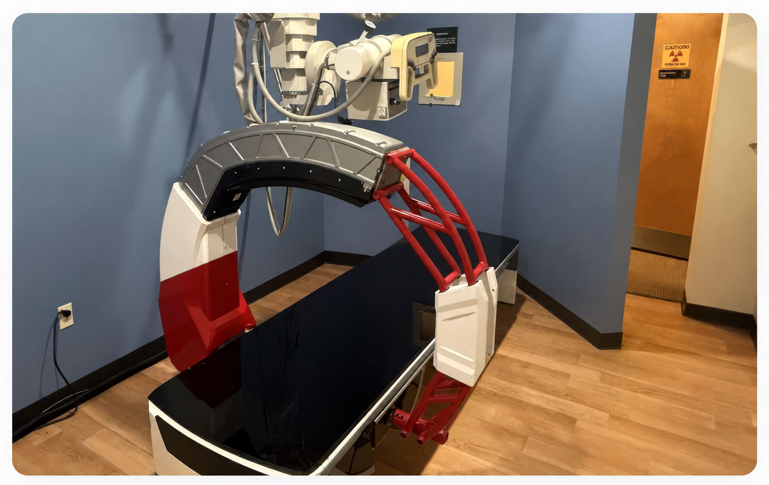
The Nanox.ARC system, installed at Access Sports Medicine.
For orthopedic practices, an X-ray doesn’t always provide all the information needed to answer the clinical question, often leading to a referral for additional imaging.
Bringing 3D imaging in-house offers another option — but raises important questions: What does it take to implement? Will existing infrastructure support it? How does reimbursement work? And will it deliver meaningful value for clinicians, staff, and patients?
Access Sports Medicine, an orthopedic and sports medicine group with multiple locations in southern New Hampshire, worked through those questions when it implemented Nanox.ARC, an FDA-cleared imaging technology that produces 3D tomographic images.
Its experience offers practical lessons for other orthopedic practices considering in-house 3D imaging.
Why Access Sports Medicine Considered In-House 3D Imaging
For Access Sports Medicine, the decision to explore 3D imaging started with patient care. The practice wanted to give providers another imaging option when an X-ray didn’t fully answer the clinical question, while reducing unnecessary referrals outside the practice for additional imaging.
For Greg Wainwright, Director of Diagnostic Imaging, the next question was whether that clinical opportunity could work practically within the existing practice. “When someone in my position is evaluating a new piece of equipment, you generally have a short list of questions: what does it take to operate, what does reimbursement look like, and is this going to be a headache for my team.”
— Greg Wainwright, Director Diagnostic Imaging, Access Sports Medicine
Those questions are particularly important when a practice already has established X-ray workflows, technologists, imaging infrastructure, and referral relationships.
The goal isn’t necessarily to replace what already works. It’s to determine whether an additional imaging capability can fit into the existing care pathway and provide useful information in cases where conventional radiography may not tell the entire story.
For Access Sports Medicine, that evaluation led to Nanox.ARC.
What Is Nanox.ARC 3D Imaging?
Nanox.ARC is an FDA-cleared 3D imaging system intended to produce 3D tomographic images.
Using digital tomosynthesis, the system acquires X-ray images from multiple angles and reconstructs them into a series of tomographic images. A single scan can generate up to 100 coronal views, providing clinicians with additional visualization of anatomy beyond a single planar X-ray image.
Nanox.ARC is cleared for general use, including musculoskeletal, pulmonary, intra-abdominal, and paranasal sinus indications, adjunctive to conventional radiography, on adult patients in professional healthcare settings such as hospitals, clinics, and imaging centers. For equivalent body parts, Nanox.ARC uses up to 80% less radiation than CT.
For an orthopedic practice like Access Sports Medicine, the question was how that capability would translate into day-to-day operations and clinical care.
What Does It Take to Add 3D Imaging to an Orthopedic Practice?
One of Access Sports Medicine’s biggest lessons was that implementing Nanox.ARC required less infrastructure change than the team initially anticipated.
Using an Existing X-Ray Room
Nanox.ARC can fit in a standard shielded X-ray room as small as 15 × 12 feet and operates using a standard power outlet.
At Access Sports Medicine, the system was installed in an existing X-ray room without major construction or specialized electrical requirements.
Wainwright recalled a representative from a large imaging equipment manufacturer visiting the practice after installation and asking a simple question:
“Does this thing really just plug into a wall?”
Wainwright’s answer:
“Absolutely. We didn’t do any construction. We didn’t need any special power requirements. It went right into our existing X-ray room.”
— Greg Wainwright
For practices evaluating 3D imaging, understanding whether existing infrastructure can be an important part of assessing implementation requirements, potential downtime, and cost.
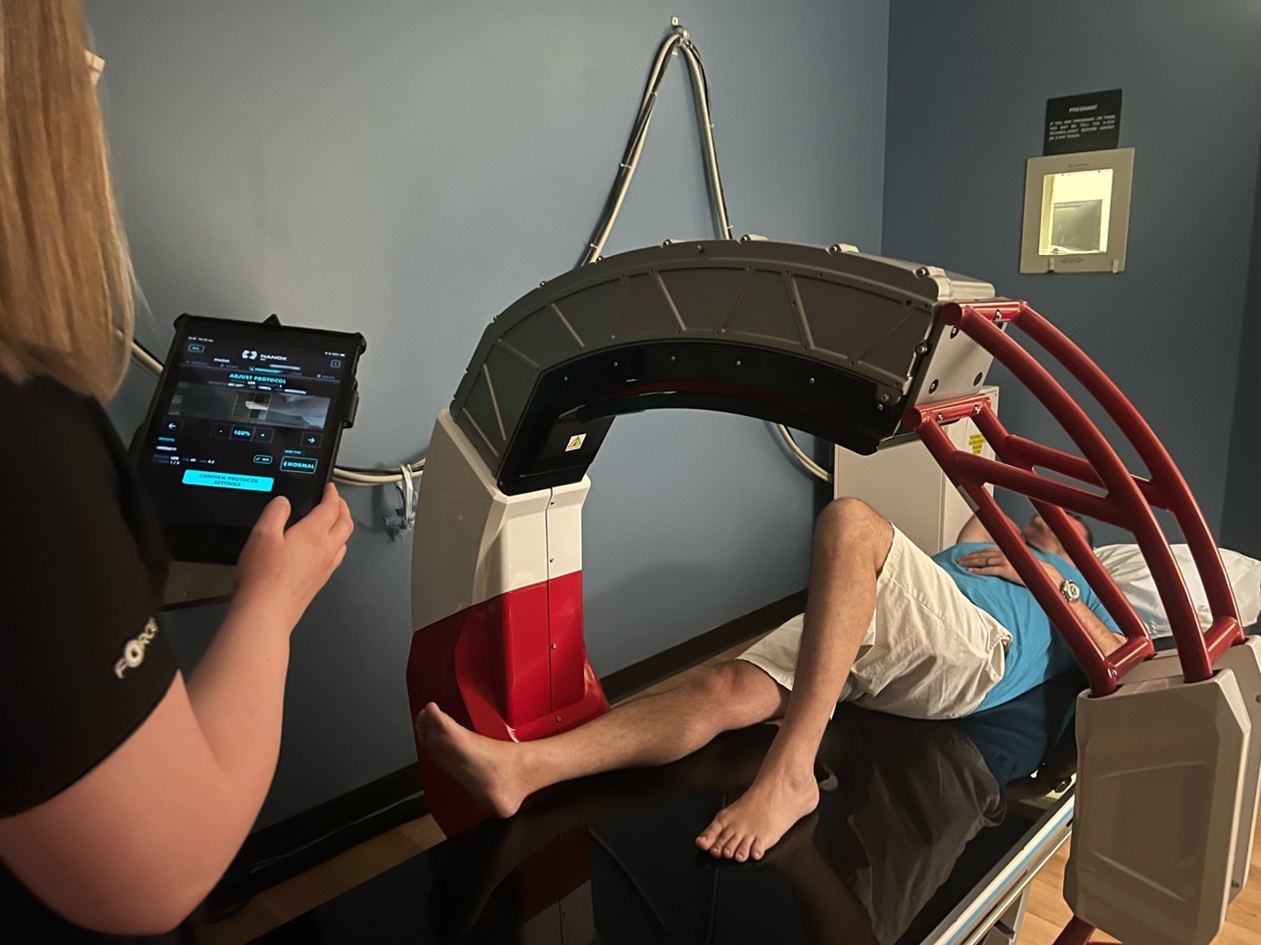
A technologist at Access Sports Medicine uses the interface to prepare a patient for a scan, which lasts a few seconds
Integrating 3D Imaging Into the Existing Workflow
Adding a new imaging technology also raises questions about staffing and training.
According to Wainwright, Access Sports Medicine’s existing radiologic technologists adapted quickly to Nanox.ARC, and the practice did not need to hire specialized staff to operate the system.
“The ARC can be operated by an X-ray technologist—it doesn’t require bringing in a CT technologist or adding a new position.”
That allowed the practice to incorporate 3D imaging into its existing radiology workflow rather than building an entirely new operational process around the technology.
Initial training helped the team incorporate Nanox.ARC into its existing radiology workflow, while ongoing education and support continue to help technologists refine positioning, build confidence, and apply what they learn across different clinical scenarios. For Access Sports Medicine, that continued training has been an important part of getting the most value from the technology over time.
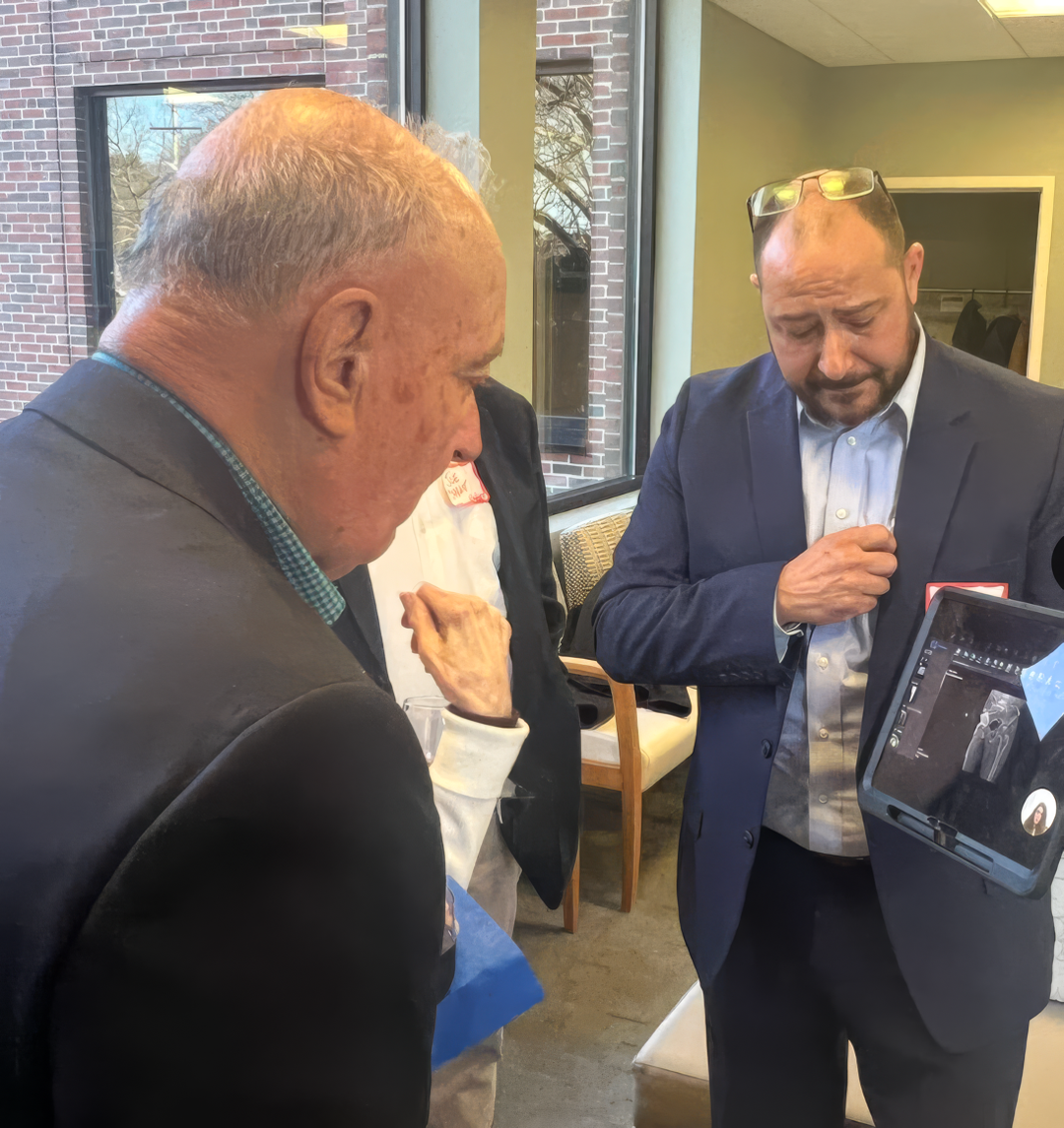
Matt Cohen, Clinical Education Specialist with Nanox, illustrates the clinical value of the Nanox.ARC during a launch event at Access Sports Medicine
Planning for PACS and Image Storage
One implementation consideration was less obvious: image storage.
Because a Nanox.ARC scan can produce many more images than a conventional X-ray exam, practices should involve their PACS and IT teams early in the planning process.
Access Sports Medicine found that storage requirements can resemble those associated with larger imaging studies more than traditional X-ray exams.
For practices considering implementation, Wainwright’s experience points to a simple lesson: don’t treat PACS capacity as an afterthought.
Discuss expected study size, image transfer, storage capacity, reading workflows, and system compatibility before go-live and loop in your PACS administrator early.
How Is Nanox.ARC Reimbursed?
Reimbursement was another important consideration for Access Sports Medicine.
Nanox.ARC exams are typically billed under CPT 76100. Coverage, coding, reimbursement, and prior authorization requirements can vary by payer, plan, and patient, so practices should verify requirements with the applicable insurer.
For Wainwright, having an established billing pathway helped make the business discussion more straightforward.
“Exams are typically billed under CPT 76100. That clarity alone made the conversation with our executive team easy.”
— Greg Wainwright
Practices should conduct their own reimbursement analysis based on their payer mix, contracts, and market before implementing a new imaging service.
Where Access Sports Medicine Sees Clinical Value
Operational simplicity only matters if the technology also provides clinical value.
Dr. David Davis, the practice’s Managing Partner, sees Nanox.ARC as another step in the imaging pathway, giving clinicians access to more anatomical detail than a conventional X-ray when the clinical question may not yet warrant a CT.
“What I like about Nanox.ARC is that it gives us another level of information when an X-ray doesn’t give us everything we need. There are cases where we want a better look at the anatomy, but the patient may not need to go straight to CT. This gives us another option in that decision-making process.”
— Dr. A, David Davis, Managing Partner,, Access Sports Medicine
Nanox.ARC does not replace conventional X-ray or CT. It provides a 3D imaging option clinicians can consider based on the individual patient and clinical questions.
For Access Sports Medicine, that has created an additional imaging capability within the practice itself.
A Patient Case: When the Initial X-Ray Didn’t Provide a Clear Answer
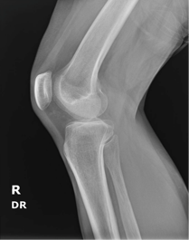
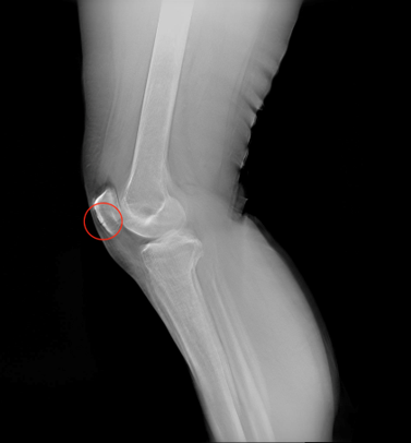
One patient came to Access Sports Medicine with knee pain after previously receiving an X-ray at an urgent care clinic that had not shown clear findings.
Because the Nanox.ARC does not require pre-authorization, Access Sports Medicine was able to perform a Nanox.ARC scan the same day.
The resulting tomographic images provided the clinical team with additional visualization to factor into its assessment. Based on the complete clinical evaluation, the patient was referred for appropriate follow-up care as part of continued treatment.
The case illustrates where Access Sports Medicine sees a potential role for in-office 3D imaging: providing additional information when conventional radiography does not fully answer the clinical question, without automatically requiring the patient to leave the practice for another imaging appointment.
X-ray / Nanox.ARC comparison image of a patella
Three Lessons From Implementing In-Office 3D Imaging
Looking back on implementation, Access Sports Medicine’s experience highlights several considerations for other orthopedic practices evaluating 3D imaging.
1. Existing Imaging Infrastructure May Be Enough
Adding an imaging modality does not always mean building a new imaging suite. Nanox.ARC was installed in Access Sports Medicine’s existing shielded X-ray room without any construction or specialized electrical requirements.
2. Involve PACS and IT Early
A 3D imaging study generates substantially more images than a conventional X-ray. Practices should plan for image transfer, PACS integration, storage, and reading workflows before installation.
3. Consider Workflow and Training, Not Just Equipment
A new imaging system has to work for the people operating it. Access Sports Medicine was able to train its existing radiologic technologists rather than hiring specialized new staff, and ongoing training has remained an important part of building confidence, refining technique, and getting the most value from 3D imaging over time. Plan time for training and get your technologists involved early.
For Wainwright, the operational and clinical experience has ultimately been positive.
“For us, Nanox.ARC has been a win operationally and clinically. Honestly, my only regret is not putting it in earlier.”
— Greg Wainwright
Frequently Asked Questions About Nanox.ARC and In-Office 3D Imaging
What does it take to install Nanox.ARC in an orthopedic practice?
Nanox.ARC fits in a standard shielded X-ray room as small as 15 × 12 feet and operates using a standard power outlet. Depending on a practice’s existing infrastructure and applicable requirements, it is often possible to install the system in an existing X-ray room without any construction.
How is Nanox.ARC billed?
Nanox.ARC exams are typically billed under CPT code 76100. Coverage, coding, reimbursement, and prior authorization requirements vary by payer and plan and should be verified for each applicable patient and insurer. However, pre-authorization is typically not required when working with in-network providers.
How much radiation does Nanox.ARC use compared with CT?
Nanox.ARC uses up to 80% less radiation than CT for equivalent body parts.
What clinical areas is Nanox.ARC FDA-cleared for?
Nanox.ARC is cleared for general use, including musculoskeletal, pulmonary, intra-abdominal, and paranasal sinus indications, adjunctive to conventional radiography, on adult patients in professional healthcare settings such as hospitals, clinics, and imaging centers.
Can existing X-ray technologists operate Nanox.ARC?
Trained radiologic technologists can operate the Nanox.ARC. Technologist training is provided by Nanox; training and staffing needs may vary by facility.
Does Nanox.ARC require a new X-ray room?
Not necessarily. Nanox.ARC can be installed in an existing shielded X-ray room, as small as 15 × 12 feet, without major construction or specialized electrical requirements.
Considering In-House 3D Imaging for Your Orthopedic Practice?
Every orthopedic practice has different clinical needs, imaging volumes, workflows, payer mixes, and infrastructure.
Access Sports Medicine’s experience demonstrates some of the practical questions worth asking when evaluating in-office 3D imaging, from room requirements and PACS planning to staff adoption, reimbursement, and clinical utility.
Explore clinical case examples and peer-reviewed research at nanox.vision/clinical-content, or contact Nanox to learn more about bringing Nanox.ARC to your practice.


 Client portal