Clinical Benefits
Enhancing Patient Care and Outcomes with Advanced Technology

for equivalent body parts

X-rays


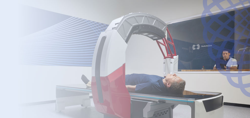
Nanox.ARC:
The Next Generation of Medical Imaging


Clinical Samples
Tomosynthesis improves clarity by reducing object superposition and minimizing pseudo-objects
 Nanox.ARC
Nanox.ARC
Nanox.ARC
Nanox.ARC
Nanox.ARC
Nanox.ARC
Nanox.ARC
Nanox.ARC
Nanox.ARC
Nanox.ARC
Nanox.ARC
Nanox.ARC
Nanox.ARC
Advanced Imaging Solution for All


Real Life Evidence
Financial Benefits
Maximize financial gains with our tailored solutions

fully integrated within the Nanox ecosystem


higher reimbursement on top of
radiography, CPT code 76100


Flexible Financing For Quick Setup
MSaaS Option:
- Effective workflow integration program.
- No capital expenditure, pay-per-scan.
- Ongoing services, maintenance, updates, and upgrades for a fee.
Discover the true value of your investment with a click
Capital Investment Option:
- One-time upfront purchase with an ongoing maintenance contract.
- Allows benefit from full ownership.
- Ongoing services, maintenance, updates, and upgrades for a fee.
Want to know more about Nanox.ARC?
| Discover the true value of your investment with a click | Calculate your ROI |
| Want to know more about Nanox.ARC? | Contact Us |
Tomosynthesis
Place in Medical Imaging
Multiple Modality Thoracic Imaging Comparison
 Anatomical Separation:
 Radiation Exposure:
 Images Generated:
 Dose:
Dose: Interpretation Time:
Explore Insights & Innovations
Explore leading articles in the field of medical imaging, featuring cutting-edge research, breakthrough technologies,
and in-depth analyses of the future of medicine. Read more and stay updated with the latest knowledge and developments.
 White Papers
 Evaluation Of The Diagnostic Potential Of A Novel Tomosynthesis System For MSK
Digital tomosynthesis (DTS) is a well-established technology that has become the gold standard for breast mammography. In recent years its benefits in musculoskeletal (MSK) imaging have been acknowledged and lead to a rapid increase in its utilization.
 5 Minutes reading
 Media
Media
COLD CATHODE DIGITAL TOMOSYNTHESIS: The evolution of high resolution, low dose imaging
COLD CATHODE DIGITAL TOMOSYNTHESIS: The evolution of high resolution, low dose imaging.
By Gregory Kicska, MD, PhD Cardiothoracic Imaging Associate Professor, University of Washington Veterans Health Administration, Seattle
White Papers Evaluation Of The Diagnostic Potential Of A Novel Tomosynthesis System For Chest And Lung Diseases
Chest Digital Tomosynthesis (DTS) is known for its capability to overcome significant chest radiographs limitations (CXR). It improves detection of chest lesions and their anatomic localization and can exclude false lesions.
5 Minutes reading

Nanox.ARC Technology
Nanox.ARC: Digital multi-source 3D tomosynthesis imaging system, globally integrated for advanced radiographic visualizations

Nanox.SOURCE
A novel silicon-based, low voltage, nano-scale cold cathode generating the electron stream needed for X-ray via field-emission technology
Read More about Nanox.SOURCE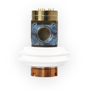

Nanox.CLOUD
Digital image processing
A secure, proprietary service for image processing, which can be hosted locally or cloud based
Nanox.AI
We are developing a Pulmonary solution which is expected to be the first AI application designed specifically for the Nanox.ARC
Read More about Nanox.AI


 Client portal