Advanced 3D imaging that was built for access and powered by innovation
Introducing Nanox.ARC X


From Rare to Everywhere. Bringing advanced 3D imaging to every point of care.
Nanox.ARC X: Where Less Delivers More


Advanced Imaging Solution for All


Urgent Care Centers
Chiropractors
Pain Management
Sports Medicine
Designed for access today
Ready for the intelligence of tomorrow
Multiple Modality Thoracic Imaging Comparison
 Image Quality:
overlapping body structures
 Radiation Dose:
0.04 – 0.1 mSv
0.1 – 0.2 mSv
Low Dose CT: 1 to 1.5 mSv
 Images Generated:
 Interpretation Time:
Financial Benefits
Maximize financial gains with our tailored solutions

fully integrated within the Nanox ecosystem

capital purchase

higher reimbursement on top of
radiography, CPT code 76100

comprehensive and faster clinical care

comprehensive and faster clinical care
Flexible Financing For Quick Setup
Msaas Model:
- Effective workflow integration program.
- No capital expenditure, $30 per scan (subject to minimum scans per day).
- Ongoing services, maintenance, updates, and upgrades for a fee.
Want to know more about the ARC X?
Capital Investment Option:
- One-time upfront purchase with ongoing maintenance contract.
- Allows benefit from full ownership.
- Ongoing services, maintenance, updates, and upgrades for a fee.
| Want to know more about the ARC X? | Contact Us |
Nanox.ARC Case Review
Study of a Human Foot displaying no superimpositions of anatomic structures
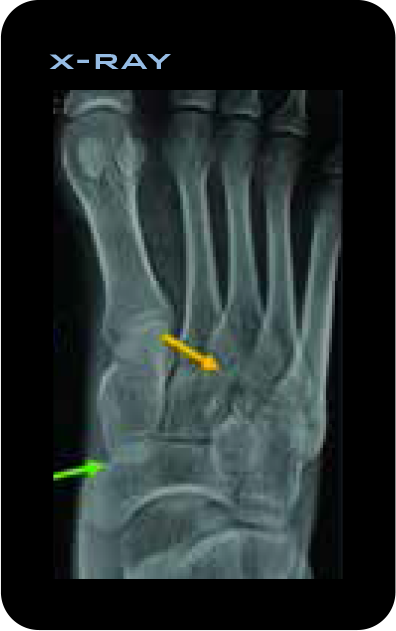
On the standard X-ray there is a line that can be suspected of being a nondisplaced fracture. The base of the medial cuneiform is obscured by the navicular on the X-ray and is better seen on Nanox.ARC.
On Nanox.ARC tomosynthesis images, the line clearly represents the cortex of the lateral cuneiform. The 4th tarsometatarsal joint is better seen in the Nanox.ARC - image (red arrow) compared to conventional radiograph.
Study of a Human Pelvis and identification of a sclerotic lesion
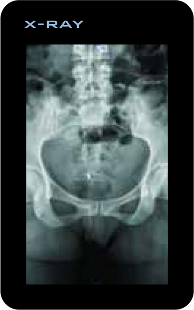
On the standard X-ray with a cast, there is a limited evaluation of the fracture and of the anatomy. The fracture is barely seen and cannot be characterized.
A sclerotic lesion in the posterior aspect of the left ramus pubis is seen using Nanox.ARC (red arrow) and not seen on the standard X-ray.
Study of a Wrist Fracture

The lesion is not seen on a standard X-ray.
On Nanox.ARC tomosynthesis images, the fracture is well seen and characterized.
Study of a Thoracic Skeleton

The fracture is not seen on the standard X-ray.
A subtle nondisplaced rib fracture is seen using the Nanox.ARC. Anatomy prevents visualization on the standard X-rays. Fracture was later confirmed on a CT Scan.
Exploring Insights and Innovations – Nanox.ARC
Explore leading articles in the field of medical imaging, featuring cutting-edge research, breakthrough technologies,
and in-depth analyses of the future of medicine. Read more and stay updated with the latest knowledge and development.
 Media
Media  Events
 ARC Day Live Broadcast
NOV 16, 2024 AT 9:30 AM EST / Neve-Ilan, Israel
Streaming live from our headquarters in Israel, we will present a demonstration of the Nanox 3D medical imaging ecosystem and discuss our commitment to improving...
 White Papers
 Evaluation Of The Diagnostic Potential Of A Novel Tomosynthesis System For MSK
Digital tomosynthesis (DTS) is a well-established technology that has become the gold standard for breast mammography. In recent years its benefits in musculoskeletal (MSK) imaging have been acknowledged and lead to a rapid increase in its utilization.
 5 Minutes reading

Nanox.ARC X Core Technologies
Nanox.ARC X: Easy-to-use tomosynthesis system, globally integrated for advanced radiographic visualizations.
Nanox.SOURCE
A novel silicon-based, low voltage, nano-scale cold cathode generating the electron stream needed for X-ray via field-emission technology
Read More about Nanox.SOURCE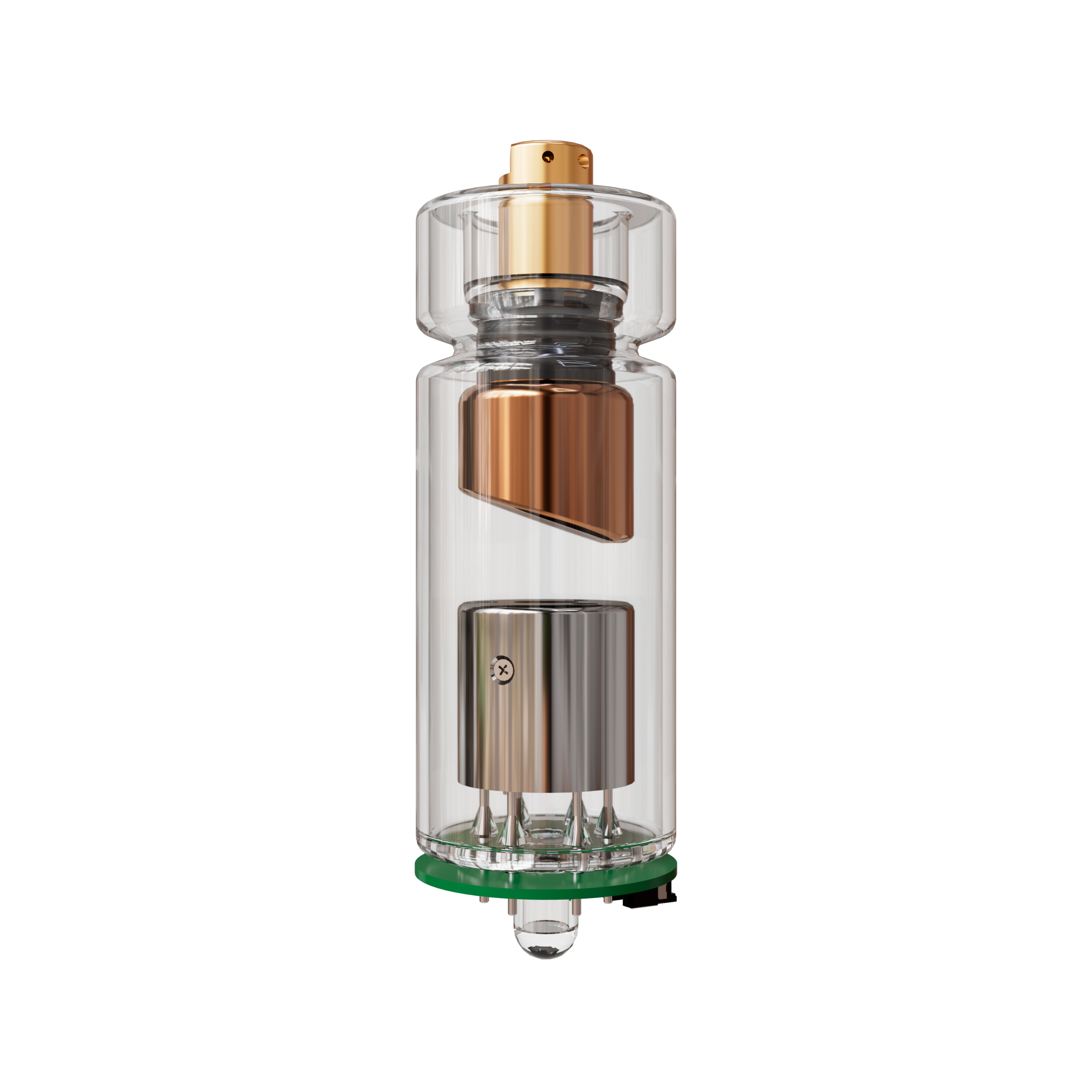

Nanox.CLOUD
Digital image processing
A secure, proprietary service for image processing, which can be hosted locally or cloud based
Nanox.AI
We are developing a Pulmonary solution which is expected to be the first AI application designed specifically for the Nanox.ARC X
Read More about Nanox.AI


 Client portal