



Introduction
In frontline radiology, the conventional 2D chest X-ray (CXR) is the absolute standard for initial lung assessment. However, architectural superimposition—where ribs, vessels, and tissues overlap—frequently masks serious conditions or creates misleading pseudo-lesions. When faced with these doubtful findings, clinical workflows traditionally mandate an escalation to Computed Tomography (CT). While highly accurate, CT scans introduce heavy operational bottlenecks, higher institutional costs, and a significantly larger patient radiation burden.
Data presented at ECR 2026 by Prof. Emilio Quaia highlights how Nanox’s cold-cathode 3D DTS system effectively eliminates this diagnostic gap. By capturing up to 100 low-dose cross-sectional slices, DTS delivers immediate diagnostic clarity, allowing clinicians to confidently rule out or confirm pulmonary pathology without leaving the X-ray suite.
Key Takeaways
Study Overview
Study Overview: Evaluating the Diagnostic Impact and Application of Chest DTS
The clinical presentation led by Prof. Emilio Quaia evaluated the definitive diagnostic utility of Digital Tomosynthesis across several critical frontline lung applications.
- Clinical Applications Evaluated: The study focused on utilizing DTS to identify and characterize pulmonary pseudolesions, lung nodule detection, lung opacities, occult lung apex lesions, and subtle chest wall alterations.
- Method & Metrics: Patient cohorts with inconclusive initial findings were scanned using both conventional chest radiography (CXR) and Digital Tomosynthesis (DTS) to measure differences in diagnostic sensitivity, specificity, interpretation time, and downstream medical costs.
- Key Metric Findings: While conventional X-ray interpretation was faster (mean of 120 seconds), it left high rates of ambiguity. Radiologists spending a mean of 200 seconds reviewing the expanded multi-slice DTS series attained near-perfect diagnostic confidence (AUC metrics ranging from 0.979 to 0.984)
Impact / Value
Why This Matters: Eliminating Unnecessary Escalations and Systemic Healthcare Costs
Integrating DTS into primary chest imaging workflows delivers immediate, measurable value to hospital administration, radiological staff,
and patient care models alike.
Real-World Impact Highlights:
- Immediate Problem Resolution: Instead of putting patients on long waiting lists for a secondary CT scan, doubtful lesions are solved immediately inside the primary X-ray unit.
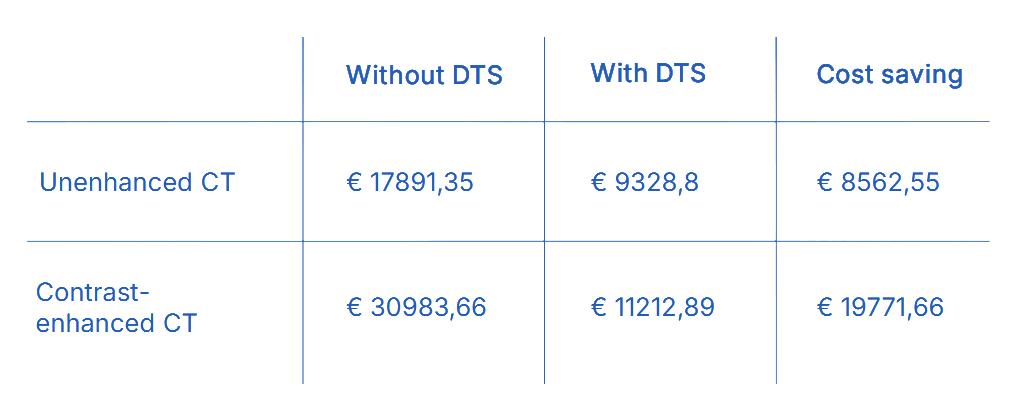
- Profound System Cost Savings: Economic modeling tracks a massive reduction in annual expenditures when replacing routine inconclusive CT paths with a localized DTS workflow. Annual costs for unenhanced CT imaging dropped from €17,891.35 to €9,328.80 (saving €8,562.55), while contrast-enhanced CT costs dropped from €30,983.66 to €11,212.89—yielding a net cost saving of €19,771.66.
- Optimized Equipment Allocation: Clearing 3/4 of doubtful cases via tomosynthesis directly prevents specialized CT machinery from being overloaded with basic diagnostic rule-out tasks.
Frequently Asked Questions
Digital Tomosynthesis is highly effective at ruling out pulmonary pseudolesions caused by overlapping tissue, detecting early lung nodules, defining vague lung opacities, revealing hidden lung apex lesions, and evaluating structural chest wall alterations.
DTS represents a highly optimized low-dose alternative. While a standard diagnostic chest CT scan carries an estimated radiation exposure of 1 to 3 mSv, a comprehensive chest DTS scan exposes the patient to a mean dose of only 0.2 mSv.
By preventing unnecessary follow-up CT scans, a DTS-supported workflow dramatically reduces institutional spending. Clinical cost matrices demonstrate annual savings of up to €8,562 for unenhanced chest imaging pathways and over €19,771 for contrast-enhanced clinical tracks.
Related Resources
Nanox.ARC: Next-Generation Multi-Source 3D Tomosynthesis Imaging
Activating Latent Data: The Evolving Role of AI in CT Workflows
Evaluation of the Diagnostic Potential of Novel Tomosynthesis in MSK Workflows

Full Professor of Radiology & Chair – University of Padova (Italy) Prof. Quaia is a world-renowned radiologist and the Director of Radiology at the University of Padova, as well as an Honorary Clinical Senior Lecturer at the University of Edinburgh. A leading pioneer in chest imaging and advanced digital tomosynthesis applications, his clinical workflows focus on optimizing diagnostic precision and lowering patient radiation exposure.


 Portail client